The psoas, iliacus, and some hip release options
I have dedicated the last three months of reading to the pelvis, specifically to the psoas major. This happens; I read something about a technique or body part, and then I decide to see how many books exist on the subject that I can easily acquire. (My husband no longer rolls his eyes when he sees an Amazon box filled with 4 books on the same anatomical/movement subject by different authors. Acceptance is a foundation for all relationships). The psoas major is interesting. It stabilizes the spine, connects the lower body to the torso, and has several different actions, depending on the movement occurring. Dr. Kathy Dooley wrote a great blog on the anatomy and stretching the psoas here.
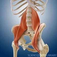
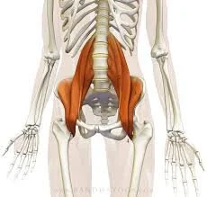
The thing that has always struck me about the psoas major is how big it is. When you hang out in an anatomy lab, this muscle is much meatier than you might expect. It’s attachment points are the discs from T12-L5, the transverse processes from L1-L5, and the lessor trochanter of the femur (Koch, 2012, Bowman, 2014). This means the psoas major starts at the bottom of the ribcage and attaches on your lower leg bone. That’s impressive. If you imagine (or look at the picture) pulling on the attachment point at the femur, the psoas major will flex the leg; however, if you imagine pulling on the attachment points on the vertebrae, the trunk will flex or extend, depending on where/how you pull. According to Koch, 2012, a healthy psoas isn’t a strong flexor of the hip during walking. Rather, it functions to keep the spine in a state of neutrality in the frontal and transverse planes, allowing the leg to move in a variety of ways without pulling on the core. This is in juxtaposition to the iliacus, which has attachment points on the iliac bone of the pelvis and the lesser trochanter of the femur, putting in a far more optimal position to flex the hip (Staugaard-Jones, 2012). In a supine position, the psoas acts to compress the lumbar spine during spinal flexion, specifically during a sit-up or roll-up motion. The psoas has fascial connections with the diaphragm, making it’s resting position dependent upon the position of the diaphragm. When the hips are in a chronically shortened position, this affects resting psoas length and can alter not just hip extension, but rib cage and neck position as a means of compensation, making the psoas a “neurological driver” for the rest of the body’s alignment (Bowman, 2014) . A chronically shortened psoas commonly looks like a posterior pelvic tilt with rib thrusting, while a chronically shortened iliacus usually looks like an anterior pelvic tilt. There are always exceptions, of course, but when you look at the position of the muscles, anatomically, this should make sense.

The next logical question is what can be done to improve alignment of these muscles? Obeying rib position is key for improving resting length of either the psoas major or the iliacus. Ribs that are thrusted forward are compressing in the back body and will affect diaphragm position and function, so ensuring an expiratory, or “neutral” rib position is key. If the individual presents with a shortened psoas, I like the psoas release that Katy Bowman describes both in her book “Move your DNA,” and in her lecture “The Science of the Psoas.” Sit with your legs in front of you, thighs in contact with the floor. Place a bolster behind you, length-wise, and lie back on it. The bolster position will be determined by whether or not the back of the thighs stay on the ground. If the thighs come off the ground, move the bolster closer to the pelvis. Stay here for a few minutes (5 or so), and allow everything to release. Exercises performed in 1/2 kneeling can be powerful, with alignment being paramount. If the right leg is forward, make sure the hips are even (the right side will have a tendency to hike up, so make sure it’s level with the left). From here, I often give the cue, “lift the right lower belly away from the right thigh.” This will get the spine out of flexion and put everything in a more balanced position. There is a dynamic exercise I demonstrate below that has worked well for me. Finally, if anterior pelvic tilt is present and I want to release some of the back muscles and put the iliacus in a more optimal position, I use Eric Franklin’s ball exercise. In a supine position with the knees bent, feet flat on the floor, place a ball under each pelvis (I have used both the Franklin balls for this and the Yoga Tune Up balls, and liked both. Tennis balls would probably work as well, though they might be a little bit small). Pick one foot up, lower it done, and then the other foot up, lower it down, marching in place from the hip. You should feel a sense of ease during hip flexion, and when you remove the balls and lie on the ground, you might notice your back muscles are more relaxed. Breathing exercises can be done in conjunction with the releasing exercises. It is important to remember movement inefficiencies can be driven by issues in the eyes, jaw, or feet, not just in the core. Psoas or iliacus tightness can also be asymmetrical, so just because it shows up on one side, it doesn’t guarantee it will show up on the other.
It is worthwhile to note the psoas is considered an emotional center in Eastern medicine, and is thought to be a place people carry stress. Suggested reading on this topic can be found in the reference section below, but noticing how you clench when stress is present (and when it’s not) can have a profound affect on overall well-being.
Yours in health and wellness,
Jenn
Dynamic hip flexor stretch:
https://www.youtube.com/watch?v=GFm9z75tDUY
References:
Koch, L., (2012). Core Awareness. Berkeley: North Atlantic Books.
Staugaard-Jones, JA., (2012). The Vital Psoas Muscle: connecting physical, emotional, and spiritual well-being. Berkeley: North Atlantic Books
Bowman, K., (2014). The Science of the Psoas.